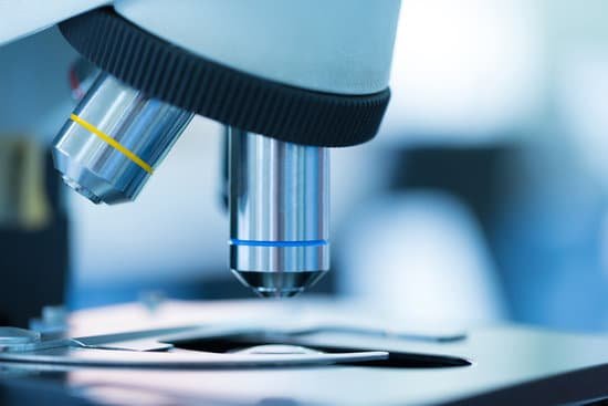
Who is credited with first observing cells in a microscope? Robert Hooke was the first to use a microscope to observe living things. Hooke’s 1665 book, Micrographia, contained descriptions of plant cells.
Can we see DNA in microscope? Yes, but not in detail. “Many scientists use electron, scanning tunneling and atomic force microscopes to view individual DNA molecules,” said Michael W. Davidson, curator of the National High Magnetic Field Laboratory at Florida State University.
What type of microscope can you see DNA? Electron microscopes, too, can see DNA in cells, and DNA sequencers can determine the A’s, T’s, C’s, and G’s (nucleotides) it’s made of.
What magnification do you need to see DNA? A stain has been used to dye DNA in the cells. This will make the chromosomes show up in a darker color than the rest of the cell. Focusing the microscope with 40x objective should give you a close enough view of the chromosomes to find each phase.
Who is credited with first observing cells in a microscope? – Related Questions
Are there microscopic organisms in clouds?
When you look at a cloud, you’re actually looking at billions of microorganisms. … Scientists have identified thousands of species of microbes and fungi living in rain and clouds—able to withstand the harsh temperatures, low oxygen and punishing radiation of the atmosphere.
What does condenser do in microscope?
On upright microscopes, the condenser is located beneath the stage and serves to gather wavefronts from the microscope light source and concentrate them into a cone of light that illuminates the specimen with uniform intensity over the entire viewfield.
Can particles in a suspension be seen through a microscope?
The internal phase (solid) is dispersed throughout the external phase (fluid) through mechanical agitation, with the use of certain excipients or suspending agents. An example of a suspension would be sand in water. The suspended particles are visible under a microscope and will settle over time if left undisturbed.
How was the first compound microscope different from leeuwenhoek?
Whereas van Leeuwenhoek used a simple microscope, in which light is passed through just one lens, Galileo’s compound microscope was more sophisticated, passing light through two sets of lenses.
What type of microscope do you need for bacteria?
In order to actually see bacteria swimming, you’ll need a lens with at least a 400x magnification. A 1000x magnification can show bacteria in stunning detail. However, at a higher magnification, it can be increasingly difficult to keep them in focus as they move.
How to take microscope pictures with iphone?
Open the application and focus the object correctly in the microscope. Bring the camera in the phone near the eye piece and click a photo once you get the object correctly focused. Hit ‘Use’ and put in the magnification of the image. Hit ‘Accept’ and view the image.
What type of microscope is used to view discover cells?
Two types of electron microscopy—transmission and scanning—are widely used to study cells. In principle, transmission electron microscopy is similar to the observation of stained cells with the bright-field light microscope.
Which scientist perfected the microscope?
In the late 16th century several Dutch lens makers designed devices that magnified objects, but in 1609 Galileo Galilei perfected the first device known as a microscope.
What kind of microscope uses visible light?
The optical microscope, often referred to as the “light optical microscope,” is a type of microscope that uses visible light and a system of lenses to magnify images of small samples.
Are there microscopic bugs in your eyelashes?
Eyelash mites are tiny cigar-shaped bugs found in bunches at the base of your eyelashes. They’re normal and usually harmless, unless you have too many of them. Also known as demodex, each mite has four pairs of legs that make it easy to grip tube-shaped things — like your lashes.
What is microscopic bacteria commonly known as?
Technically a microorganism or microbe is an organism that is microscopic. The study of microorganisms is called microbiology. Microorganisms can be bacteria, fungi, archaea or protists. The term microorganisms does not include viruses and prions, which are generally classified as non-living.
What is the function of magnets in an electron microscope?
Electron microscopes use shaped magnetic fields to form electron optical lens systems that are analogous to the glass lenses of an optical light microscope.
How to work out the magnification of a microscope?
To calculate the total magnification of the compound light microscope multiply the magnification power of the ocular lens by the power of the objective lens. For instance, a 10x ocular and a 40x objective would have a 400x total magnification. The highest total magnification for a compound light microscope is 1000x.
What are two different types of electron microscopes?
Today there are two major types of electron microscopes used in clinical and biomedical research settings: the transmission electron microscope (TEM) and the scanning electron microscope (SEM); sometimes the TEM and SEM are combined in one instrument, the scanning transmission electron microscope (STEM):
Who used the first microscope to study cells?
Interested in learning more about the microscopic world, scientist Robert Hooke improved the design of the existing compound microscope in 1665. His microscope used three lenses and a stage light, which illuminated and enlarged the specimens.
When was microscopic colitis discovered?
Researchers (Read, et al.) used the term “microscopic colitis” in 1980 to describe the entity of chronic, watery diarrhea in patients with only microscopic evidence of inflammation. The term is currently used to include collagenous and lymphocytic colitis. What are Collagenous and Lymphocytic Colitis?
Can we see protein synthesis with a microscope?
Individual protein molecules are very difficult to observe because of their tiny size. The scientists, led by Marvin Tanenbaum, developed a special protein staining technique called ‘SunTag’, with which single proteins can be seen in real-time using a sensitive microscope while they are undergoing synthesis.
What type of microscope does not use light?
Electron microscopes differ from light microscopes in that they produce an image of a specimen by using a beam of electrons rather than a beam of light. Electrons have much a shorter wavelength than visible light, and this allows electron microscopes to produce higher-resolution images than standard light microscopes.
How to clean a microscope camera?
To clean the eyepiece lens of your microscope, breathe onto the eyepiece lens and then wipe with lens tissue. For dirt that is difficult to remove, add ethanol (methanol in extreme cases) to a cotton swab, wipe the surface and then dry with a dry swab.
How are microscopic structures different from macroscopic structures?
In the context of Chemistry, “microscopic” implies the atomic or subatomic levels which cannot be seen directly (even with a microscope!) whereas “macroscopic” implies things that we can know by direct observations of physical properties such as mass, volume, etc.

