Why is the image upside down in a microscope? Under the slide on which the object is being magnified, there is a light source that shines up and helps you to see the object better. This light is then refracted, or bent around the lens. Once it comes out of the other side, the two rays converge to make an enlarged and inverted image.
Why are images upside down and backwards in a microscope? There are also mirrors in the microscope, which cause images to appear upside down and backwards. … The letter appears upside down and backwards because of two sets of mirrors in the microscope. This means that the slide must be moved in the opposite direction that you want the image to move.
Does a microscope make an image upside down? A microscope is an instrument that magnifies an object. … The optics of a microscope’s lenses change the orientation of the image that the user sees. A specimen that is right-side up and facing right on the microscope slide will appear upside-down and facing left when viewed through a microscope, and vice versa.
What is a dissection microscope? A dissecting microscope, also known as a stereo microscope, is used to perform dissection of a specimen or sample. It simply gives the person doing the dissection a magnified, 3-dimensional view of the specimen or sample so more fine details can be visualized.
Why is the image upside down in a microscope? – Related Questions
What does the iris diaphragm do on a microscope?
Iris Diaphragm controls the amount of light reaching the specimen. It is located above the condenser and below the stage. Most high quality microscopes include an Abbe condenser with an iris diaphragm. Combined, they control both the focus and quantity of light applied to the specimen.
What is the magnification of an eyepiece microscope?
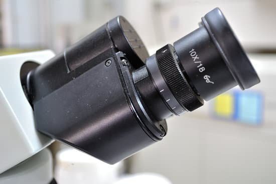
The standard eyepiece magnifies 10x. Check the objective lens of the microscope to determine the magnification, which is usually printed on the casing of the objective.
How to view fungi under the microscope?
To identify fungi under microscope the best technique is a slide culture, first from the primary plate/tube make slide culture of overnight to 48 hours.
What is meant by limit of resolution of a microscope?
The limit of resolution (or resolving power) is a measure of the ability of the objective lens to separate in the image adjacent details that are present in the object. … The resolving power of an optical system is ultimately limited by diffraction by the aperture.
What is the nosepiece used for on a microscope?
Nosepiece houses the objectives. The objectives are exposed and are mounted on a rotating turret so that different objectives can be conveniently selected. Standard objectives include 4x, 10x, 40x and 100x although different power objectives are available. Coarse and Fine Focus knobs are used to focus the microscope.
Who created the 1st microscope?
It’s not clear who invented the first microscope, but the Dutch spectacle maker Zacharias Janssen (b. 1585) is credited with making one of the earliest compound microscopes (ones that used two lenses) around 1600. The earliest microscopes could magnify an object up to 20 or 30 times its normal size.
What is coverslip in microscope?
When viewing any slide with a microscope, a small square or circle of thin glass called a coverslip is placed over the specimen. It protects the microscope and prevents the slide from drying out when it’s being examined. The coverslip is lowered gently onto the specimen using a mounted needle .
What does hep c look like under a microscope?
“It looks like a simple little white sphere among other white spheres lipid in the blood,” Meunier described. “This kind of fat sandwich is made in the center of the RNA viral and nucleus of virus separated by a first monolayer of phospholipids.
What is the path of light through a compound microscope?
The path of light through a microscope. Modern microscopes are complex precision instruments. Light, originating in the light source (1), is focused by the condensor (2) onto the specimin (3). The light then enters the objective lens (4) and the image is magnified.
What is a microscope simple definition?
A microscope is an instrument that can be used to observe small objects, even cells. The image of an object is magnified through at least one lens in the microscope. This lens bends light toward the eye and makes an object appear larger than it actually is.
How many types of microscope?
There are several different types of microscopes used in light microscopy, and the four most popular types are Compound, Stereo, Digital and the Pocket or handheld microscopes. Some types are best suited for biological applications, where others are best for classroom or personal hobby use.
Who invented the simple light microscope?
Two Dutch spectacle-makers and father-and-son team, Hans and Zacharias Janssen, create the first microscope. Robert Hooke’s famous “Micrographia” is published, which outlines Hooke’s various studies using the microscope.
Can nucleolus be seen with light microscope?
Thus, light microscopes allow one to visualize cells and their larger components such as nuclei, nucleoli, secretory granules, lysosomes, and large mitochondria. The electron microscope is necessary to see smaller organelles like ribosomes, macromolecular assemblies, and macromolecules.
How does the microscope help us today?
A microscope lets the user see the tiniest parts of our world: microbes, small structures within larger objects and even the molecules that are the building blocks of all matter. The ability to see otherwise invisible things enriches our lives on many levels.
What is the microscopic unit of fungi?
Microscopic features of fungi. Hyphae are the basic cellular unit of filamentous fungal structures. Individual hyphae are small and, with few exceptions, can be seen only after considerable magnification.
Is 40x in the microscope the red line?
The numbers in the top line are all different for the three objectives; the lower line contains numbers that are the same for all three objectives (see Figure 2-1). Figure 2-1. Note the markings and color band on each objective: red (4X), yellow (10X) and blue (40X).
Are c elegans microscopic?
C. elegans exhibits these phenomena, yet is only 1 mm long and may be handled as a microorganism—it is usually grown on petri plates seeded with bacteria. All 959 somatic cells of its transparent body are visible with a microscope, and its average life span is a mere 2-3 weeks.
How does a light optical microscope form an image?
The light microscope is an instrument for visualizing fine detail of an object. It does this by creating a magnified image through the use of a series of glass lenses, which first focus a beam of light onto or through an object, and convex objective lenses to enlarge the image formed.
What does a fine adjustment on a microscope mean?
Fine Adjustment Knob – This knob is inside the coarse adjustment knob and is used to bring the specimen into sharp focus under low power and is used for all focusing when using high power lenses. Light Source – The light source in your microscope is a lamp that you turn on and off using a switch.
What is the best resolving power of a microscope?
The greatest resolving power in optical microscopy is realized with near-ultraviolet light, the shortest effective imaging wavelength. Near-ultraviolet light is followed by blue, then green, and finally red light in the ability to resolve specimen detail.

