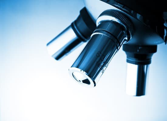
How is each optical lens used in microscope? Optical microscopes use a combination of objective and ocular lenses (eyepieces) for imaging. The observation magnification is the product of the magnifications of each of the lenses. This generally ranges from 10x to 1,000x with some models even reaching up to 2000x magnification.
What are the 4 lenses on a microscope? Magnification: Your microscope has 4 objective lenses: Scanning (4x), Low (10x), High (40x), and Oil Immersion (100x).
How are lenses used in microscope? While the modern microscope has many parts, the most important pieces are its lenses. It is through the microscope’s lenses that the image of an object can be magnified and observed in detail. … When light reflects off of an object being viewed under the microscope and passes through the lens, it bends towards the eye.
What type of lenses are used in optical microscope? Microscopes use convex lenses in order to focus light.
How is each optical lens used in microscope? – Related Questions
What microscope is best to study a membrane?
The electron microscope can achieve a much greater resolution than that obtained with the light microscope because the wavelength of electrons is shorter than that of light.
What did hooke observe about the cork under the microscope?
Hooke detailed his observations of this tiny and previously unseen world in his book, Micrographia. To him, the cork looked as if it was made of tiny pores, which he came to call “cells” because they reminded him of the cells in a monastery.
Can atom be seen under microscope?
Atoms are really small. So small, in fact, that it’s impossible to see one with the naked eye, even with the most powerful of microscopes. … Now, a photograph shows a single atom floating in an electric field, and it’s large enough to see without any kind of microscope.
What is the difference between microscopic and macroscopic ecosystems?
Microscopic approach considers the behaviour of every molecule by using statistical methods. In Macroscopic approach we are concerned with the gross or average effects of many molecules’ infractions. These effects, such as pressure and temperature, can be perceived by our senses and can be measured with instruments.
When were digital microscope invented?
The first digital Microscope was manufactured in 1986 in Tokyo, Japan which constituted a control box and a lens the was connected to the camera. This is currently known as the Hirox Co. LTD.
Did romans have microscopes?
That this occurred some 4,000 years ago in the Chow-Foo dynasty and more than 3,500 years before the “father of modern microscopy” was born is quite remarkable. … Ancient Egyptians and Romans also used various curved lenses although no reference to a compound microscope has been found.
Why does rough endoplasmic reticulum look rough under microscope?
The rough endoplasmic reticulum (ER) is called ‘rough’ because it has organelles called ribosomes attached to the surface. Ribosomes are the organelles that turn mRNA into proteins. These proteins are initially long strings of amino acids.
What uses electron microscope?
Electron microscopes are used to investigate the ultrastructure of a wide range of biological and inorganic specimens including microorganisms, cells, large molecules, biopsy samples, metals, and crystals. Industrially, electron microscopes are often used for quality control and failure analysis.
What is a coverslip on a microscope?
When viewing any slide with a microscope, a small square or circle of thin glass called a coverslip is placed over the specimen. It protects the microscope and prevents the slide from drying out when it’s being examined. The coverslip is lowered gently onto the specimen using a mounted needle .
What does bad mold look like under a microscope?
mold spores are often round, smooth, and black under the microscope. It is useful to check out black round “spores” under the microscope using top lighting in order to distinguish them from paint droplets where paint has been sprayed in the building.
Where is the eyepiece on a microscope?
Eyepiece or Ocular is what you look through at the top of the microscope. Typically, standard eyepieces have a magnifying power of 10x. Optional eyepieces of varying powers are available, typically from 5x-30x. Eyepiece Tube holds the eyepieces in place above the objective lens.
How to use a microscope condenser?
When installing the microscope condenser, rotate the coarse focus knob (1) to move the stage to its highest position. Most compound light microscopes have a small knob (2) to raise and lower the condenser holder. Lower this holder so the condenser can slide into the holder below the stage.
Can i determine my blood type with a microscope?
But if you look at a drop of your own blood under a microscope, you would see objects floating in it that look like balls and doughnuts. … Some of these objects on the surface, called antigens, can differ from person to person. These differences are what determines your blood type.
What does the scanning objective do on a microscope?
A scanning objective lens provides the lowest magnification power of all objective lenses. 4x is a common magnification for scanning objectives and, when combined with the magnification power of a 10x eyepiece lens, a 4x scanning objective lens gives a total magnification of 40x.
Why increase light when viewing a specimen under a microscope?
Changing from low power to high power increases the magnification of a specimen. The amount an image is magnified is equal to the magnification of the ocular lens, or eyepiece, multiplied by the magnification of the objective lens.
What are the 4 types of microscopes?
There are several different types of microscopes used in light microscopy, and the four most popular types are Compound, Stereo, Digital and the Pocket or handheld microscopes. Some types are best suited for biological applications, where others are best for classroom or personal hobby use.
What a microscope is made of?
A child’s microscope may have an external body shell made of plastic, but most microscopes have an body shell made of steel. If there is a mirror included, it is usually made of a strong glass such as Pyrex (a trade name for a glass made from silicon dioxide, boron dioxide, and aluminum oxide).
Where is the field number of a microscope?
Field number is the diameter of the eyepiece lens and is most often expressed in millimeters. Field of View (FOV) is the amount of the object that can be seen with a particular optic combination (eyepieces + objective lens). It is the circular area that is seen when looking through the microscope.
Why why are bacterial cells generally stained for microscopic view?
The most basic reason that cells are stained is to enhance visualization of the cell or certain cellular components under a microscope. Cells may also be stained to highlight metabolic processes or to differentiate between live and dead cells in a sample.
Is chemistry the study of submicroscopic microscopic or macroscopic?
Is Chemistry the study of the submicroscopic, the microscopic, the macroscopic, or all three? Chemistry is all of these options; by definition, Chemistry is the study of all matter and the transformations that it can undergo. Matter exists in the submicroscopic, microscopic, and the macroscopic.
What microscope gives the best resolution?
Out of all types of microscopes, the electron microscope has the greatest capability in achieving high magnification and resolution levels, enabling us to look at things right down to each individual atom.

