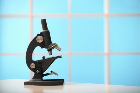
What is the function of eyepiece lens in microscope? The eyepiece, or ocular lens, is the part of the microscope that magnifies the image produced by the microscope’s objective so that it can be seen by the human eye.
What is meant by low and high power of the microscope? When you change from low power to high power on a microscope, the high-power objective lens moves directly over the specimen, and the low-power objective lens rotates away from the specimen. … The image should remain in focus if the lenses are of high quality.
What is a good power for a microscope? Most educational-quality microscopes have a 10x (10-power magnification) eyepiece and three objectives of 4x, 10x and 40x to provide magnification levels of 40x, 100x and 400x. Magnification of 400x is the minimum needed for studying cells and cell structure.
What does power mean in microscopes? Home/ Microscope Solutions/ Learn about microscope/ Resolving Power. The resolving power of an objective lens is measured by its ability to differentiate two lines or points in an object. The greater the resolving power, the smaller the minimum distance between two lines or points that can still be distinguished.
What is the function of eyepiece lens in microscope? – Related Questions
How to calculate specimen size microscope?
To estimate the size of an object seen with a microscope, first estimate what fraction of the diameter of the field of vision that the object occupies. Then multiply the diameter you calculated in micrometers by that fraction.
How to count algae cells in a microscope?
Counting of Algae count the 4 corner squares (1,3, 7 and 9; see the above figure). If the sample is very low in cells, count the whole chamber (9 squares). If you count less than 4 squares, always count the same ones, e.g. the top left and bottom right. If you count only one always count the center square.
Where does the light come from in a compound microscope?
Illuminator is the light source for a microscope, typically located in the base of the microscope. Most light microscopes use low voltage, halogen bulbs with continuous variable lighting control located within the base. Condenser is used to collect and focus the light from the illuminator on to the specimen.
Who first observed cells under a microscope?
Initially discovered by Robert Hooke in 1665, the cell has a rich and interesting history that has ultimately given way to many of today’s scientific advancements.
Are microscopic face mites harmful?
Face Mites: They Really Grow On You : Shots – Health News Demodex mites live inside your pores. Just about every adult human alive has a population living on them, and they’re basically impossible to get rid of. Luckily, they’re harmless for most people.
How to increase magnifying power of compound microscope?
From the above formula, we can conclude that the magnifying power of the compound microscope increases when the focal lengths of both objective and eyepiece lenses decrease.
When and who invented the compound microscope?
A Dutch father-son team named Hans and Zacharias Janssen invented the first so-called compound microscope in the late 16th century when they discovered that, if they put a lens at the top and bottom of a tube and looked through it, objects on the other end became magnified.
Is the image inverted in a dissecting microscope?
Binocular and dissecting microscopes will also not show an inverted image because of their increased level of magnification. … Even with an inverted image, microscopes can increase the magnification of an image phenomenally.
How do you open the iris on a microscope?
You can adjust the diaphragm by turning it clockwise to close it, or counterclockwise to open it. Only open the iris diaphragm of the microscope to a point where the light passing through barely extends beyond the microscope’s field of view.
What is an advantage of fluorescence microscope?
Fluorescence microscopy is one of the most widely used tools in biological research. This is due to its high sensitivity, specificity (ability to specifically label molecules and structures of interest), and simplicity (compared to other microscopic techniques), and it can be applied to living cells and organisms.
What is the prefix for microscope?
An easy way to remember that the prefix micro- means “small” is through the word microscope, an instrument which allows the viewer to see “small” living things.
What’s a condenser diaphragm microscope?
On upright microscopes, the condenser is located beneath the stage and serves to gather wavefronts from the microscope light source and concentrate them into a cone of light that illuminates the specimen with uniform intensity over the entire viewfield.
Does your eye move under a microscope?
When we fix our eyes on a single point, the world may appear stable, but at the microscopic level, our eyes are constantly jittering. These small eye movements, once thought to be inconsequential, are critical to the visual system in helping us reconstruct a scene, Rucci says.
What is the function of the pointer on the microscope?
Pointer: A piece of high tensile wire that sits in the eyepiece and enables a viewer to point at a specific area of a specimen.
What type of microscope is most commonly used in schools?
Compound light microscopes are one of the most familiar of the different types of microscopes as they are most often found in science and biology classrooms.
Who and when invented the microscope?
It’s not clear who invented the first microscope, but the Dutch spectacle maker Zacharias Janssen (b. 1585) is credited with making one of the earliest compound microscopes (ones that used two lenses) around 1600. The earliest microscopes could magnify an object up to 20 or 30 times its normal size.
What is the maximum resolution of an electron microscope?
The resolution limit of electron microscopes is about 0.2nm, the maximum useful magnification an electron microscope can provide is about 1,000,000x.
Can you see c elegans with a light microscope?
Visible light can be used to examine C. elegans, however, in general, bright field and phase-contrast microscopy offers little contrast- making cells and their major components difficult to see. … elegans cells (Sulston and Horvitz, 1977; Sulston et al., 1983).
What does each lens in a microscope do?
objective: the first lens light passes through after the specimen. The obective collects the light from the specimen and focusses it to a point inside the body tube. eyepiece: the lens light passes through before getting to your eye. The eyepiece magnifies the image formed by the objective so you can see your sample.
Why is there microscopic blood in my urine?
Microscopic urinary bleeding is a common symptom of glomerulonephritis, an inflammation of the kidneys’ filtering system. Glomerulonephritis may be part of a systemic disease, such as diabetes, or it can occur on its own.

